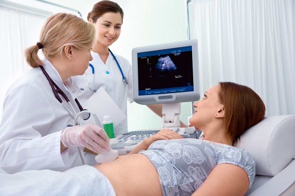
What is Ultrasonography?
■ USG is one of the first line investigation in obs. & gynecology and infertility .It can be used for inerventinal procedure also. It can be coupled with Doppler and 3D- 4D scan.
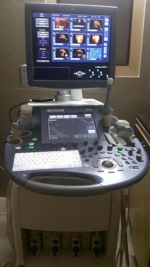
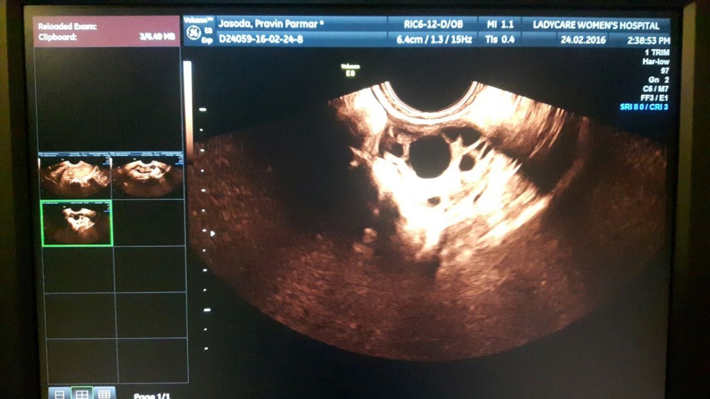
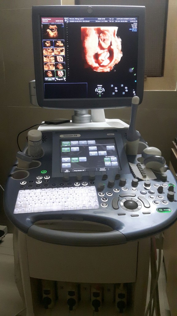
■ We have state of high tech uitrasound machine (1) Voluson E8 BTO 13.5 at clinic (2) Logiq P3
■ Ultrasonography is relatively is inexpensive, readily available , non invasive ,radiation free relatively less time consuming and easily repeatable
■ It can be performed transvaginaly and transabdomanaly
Gynecology and Infertility
■ Helps in determine the structure of uterus & fallopian tube, ovary, adnexa, blood perfusion, endometrial thickness volume & vascularity
■ It helps in detection of pathological lesion , ovarian mass, cyst, adenomyosis, endometrial fibroid, polyps etc.
■ Helps to study follicular maturity & ovulation.
■ Tubal patency can be performed by sonosalpingography
■ USG can guides oocyte retrival & embryo transfer in vitrofertilisation
■ It can guide in drainage of pelvic collection or cystic lision
■ It rules out out ectopic pregnancy
■ For gynec cancer
■ It can access pouch of doglas
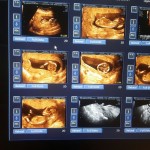
■ PICS ; Fibroid, Polyp, Ectopic, Ovarian cyst, Simple chocolate cyst
USG in obstetrics
■ Sound waves are used to create real time visual image of the developing embryo or fetus in its mother’s womb
■ It’s a standard part of antenatal clinics
■ It provides information about mother’s health, timing of pregnancy, progress of pregnancy , health and development of foetus
We recommend USG as per ISUOG:
❶ 1ST visit :conformation of pregnancy , Dating , No of foetus
❷ Between 11 to 14 wks : NT Scan screening for chromosomal abnormality and structural survey of foetus
❸ 20- 22 wks scan : for anomaly , it includes Echo
❹ Biometry doplor , echo, color Doppler
1st Trimister
✅ Early pregnancy 1st scan includes gestational sac size location and number
✅ Rule out ectopic
✅ Fetal length
✅ No of foetus , Amniotic sac, chorionic sac in case of multifoetal pregnancy
✅ Gestational sac can reliably seen on TVS at 5 wks pregnancy
11- 14 wks ( Tripple markers, Anatomical survey)
✅ Assesment of foetal age
✅ Foetal movment
✅ Placental location, cord insertion
✅ Nuchal transulancy, ductus venosus flow, & normal nasal bone helps screening chromosomal abnormality of foetus
✅ Foetal head, spine , all long bones, hands, foot, ribs.
✅ Stomach bladder , foetal kidney, genitalia and abdominal wall
✅ Foetal heart- 4 chambers out flow, ventrical septum, Aortic arch & ductal arch
✅ Cervical length
✅ If patient is found screen positive she advice for biomarker to rule out
✅ USG guided amniocentasis is done if biomarkers are abnormal
2nd Trimister
✅ Dedicated survey of extrafoetal envirment like cfoetal envirment like cervix, placental location, cord origine & insertion, amount of fluide
✅ Foetal structural biometry measurements of all bones , organ measurement , access of foetal age, maturity, growth
✅ Checks status of fetal organs it include head, spine, lungs, diaphragm, stomach, kidney, urinary blader, ant. Abdominal wall , foetal back, genitalia
✅ Detailed study of foetal heart with using advanced STIC echo
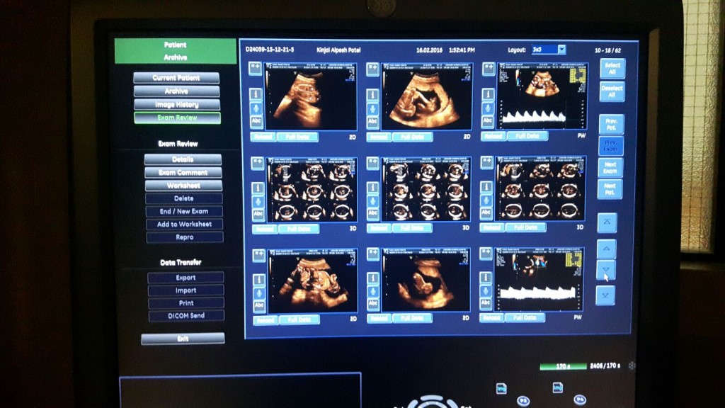
✅ Colour 3D-4D
✅ Color doplor give assessment of foetal perfusion, prediction of hypertension
✅ Assesment of risk of premature labour by messurment of cervix
✅ Soft markers give idea regarding possibility of chromosomal problems
3rd Trimister scan
✅ To see foetal position
✅ Watch for oligo
✅ Biometry to see foetal growth
✅ Doppler for foetal perfusion
✅ Echo in detail
✅ Placental maturity